Before X-rays finally reach a patient, they undergo several critical modifications to optimize their quality and minimize risk. Among these, collimation stands out as a crucial step for both image enhancement and patient safety. This article will delve into the process of collimation, explaining its mechanism, benefits, and practical application in diagnostic imaging.
The Journey of an X-ray Beam to the Patient
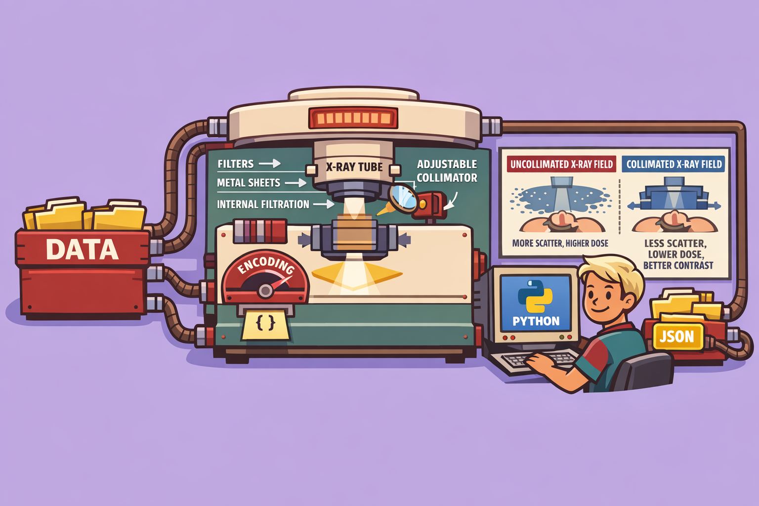
To understand collimation, let’s trace the path of an X-ray from its origin to the patient:
- X-ray Production: X-rays are initially produced isotropically (in all directions) within the X-ray tube.
- Inherent Filtration: As X-rays exit the tube, they pass through inherent filters such as the glass envelope, insulating oil, and the exit window. These materials naturally attenuate lower-energy X-rays.
- Added Filtration: Further filtering occurs with the introduction of added metal sheets, which selectively remove more low-energy photons, hardening the beam.
- Collimation: After filtration, the X-ray beam enters the collimator, the final stage before reaching the patient.
What is Collimation?
Collimation is the process of precisely shaping and restricting the X-ray beam to expose only the specific area of interest on the patient. This is achieved by using lead sheets, which effectively attenuate X-rays due to lead’s high atomic number and density, making it an excellent barrier for radiation.
How Collimation Works
Inside the collimator device, there are typically two sets of parallel lead sheets arranged perpendicularly to each other. These sheets can be moved independently to adjust the field size, creating various rectangular or square apertures. By narrowing the opening, fewer X-rays are allowed to pass through, directly reducing the irradiated area on the patient.
The Role of the Light Source:
A clever feature within the collimator is a built-in light source coupled with a mirror. This light is projected parallel to the X-ray beam, allowing radiographers to visualize the exact area that will be exposed to X-rays. This visible light field ensures accurate positioning and prevents unintentional exposure to surrounding tissues. If you’ve ever had an X-ray, you’ve likely seen this square of light on your body, marking the X-ray field.
Benefits of Collimation
Collimation offers two primary benefits:
-
Reduced Patient Dose: By limiting the X-ray beam to only the region of interest, collimation significantly decreases the total radiation dose to the patient. Non-essential areas are protected from unnecessary exposure.
-
Improved Image Quality: Collimation dramatically enhances image quality, primarily by reducing a phenomenon known as Compton scatter.
- Understanding Scatter: When X-rays interact with the patient’s body, they can undergo three main fates:
- Transmission: Pass through the patient and hit the detector.
- Photoelectric Effect: Be absorbed by the patient.
- Compton Scatter: Interact with electrons in the patient, lose some energy, and change direction. Scattered X-rays travel in random directions and can reach the detector, creating a signal that does not correspond to the actual anatomy. This “noise” reduces image contrast and resolution.
- Collimation’s Impact on Scatter: A wider, uncollimated X-ray beam exposes a larger volume of tissue, leading to more scattering events within the patient. These scattered photons can then strike the detector, degrading the image. When the beam is collimated, the volume of irradiated tissue is reduced, which consequently decreases the amount of Compton scatter generated. Less scatter means less noise on the detector, resulting in a sharper image with better contrast and spatial resolution.
- Understanding Scatter: When X-rays interact with the patient’s body, they can undergo three main fates:
It is important to note that collimation does not alter the X-ray spectrum; it doesn’t change the energy distribution of the X-rays that reach the patient, unlike filtration which removes lower-energy photons. Collimation simply removes X-rays from the periphery of the beam.
Types of Collimators
While rectangular and square collimators are most common, enabling the creation of various field sizes, other shapes exist. For specific anatomical regions or specialized X-ray procedures, circular or custom-shaped collimators can be used to further optimize radiation delivery and image capture.
Conclusion
Collimation is an indispensable tool in diagnostic radiology. It’s a win-win: radiologists achieve superior image quality with enhanced contrast and resolution, while patients benefit from significantly reduced radiation exposure. Therefore, whenever possible, the X-ray field should be tightly collimated to the area of clinical interest.
The journey of X-rays from production to image formation is complex, involving numerous physics principles. Understanding concepts like anode angle effects, the anode heel effect, filtration, and especially collimation, is fundamental to producing high-quality diagnostic images safely and effectively.
🔍 Discover Kaptan Data Solutions — your partner for medical-physics data science & QA!
We're a French startup dedicated to building innovative web applications for medical physics, and quality assurance (QA).
Our mission: provide hospitals, cancer centers and dosimetry labs with powerful, intuitive and compliant tools that streamline beam-data acquisition, analysis and reporting.
🌐 Explore all our medical-physics services and tech updates
💻 Test our ready-to-use QA dashboards online
Our expertise covers:
🔬 Patient-specific dosimetry and image QA (EPID, portal dosimetry)
📈 Statistical Process Control (SPC) & anomaly detection for beam data
🤖 Automated QA workflows with n8n + AI agents (predictive maintenance)
📑 DICOM-RT / HL7 compliant reporting and audit trails
Leveraging advanced Python analytics and n8n orchestration, we help physicists automate routine QA, detect drifts early and generate regulatory-ready PDFs in one click.
Ready to boost treatment quality and uptime? Let’s discuss your linac challenges and design a tailor-made solution!
Get in touch to discuss your specific requirements and discover how our tailor-made solutions can help you unlock the value of your data, make informed decisions, and boost operational performance!
Comments