The landscape of medicine is undergoing a profound transformation, driven by the remarkable advancements in artificial intelligence and radiomics. We are moving beyond the traditional view of medical scans as mere pictures, leveraging technology to perform what can be described as a “digital biopsy”—extracting deep biological insights from mere pixels. This shift is fundamentally changing how we understand and treat diseases, particularly in oncology.
The Digital Biopsy: Seeing Beyond the Surface
At the heart of radiomics is the understanding that machines “see” human anatomy in a fundamentally different way than humans do. For us, a brain scan reveals macroscopic shapes and contrasts. For a machine, it’s a vast grid of numbers, each representing a “voxel”—a 3D pixel. Just as a pixel is the smallest unit of a 2D image, a voxel is the foundational unit in 3D medical imaging, holding a distinct, measurable mathematical value. Whether it’s a CT scan measuring tissue density, a PET scan tracking metabolism, or an MRI mapping protons, every voxel carries a piece of a numerical puzzle.
Human eyes cannot process the ocean of invisible textural patterns and quantitative data contained within these voxels. This is where AI truly bridges the gap, allowing us to convert these everyday pixels into a virtual biological biopsy.
Inside the Radiomics Engine: The Six-Phase Blueprint
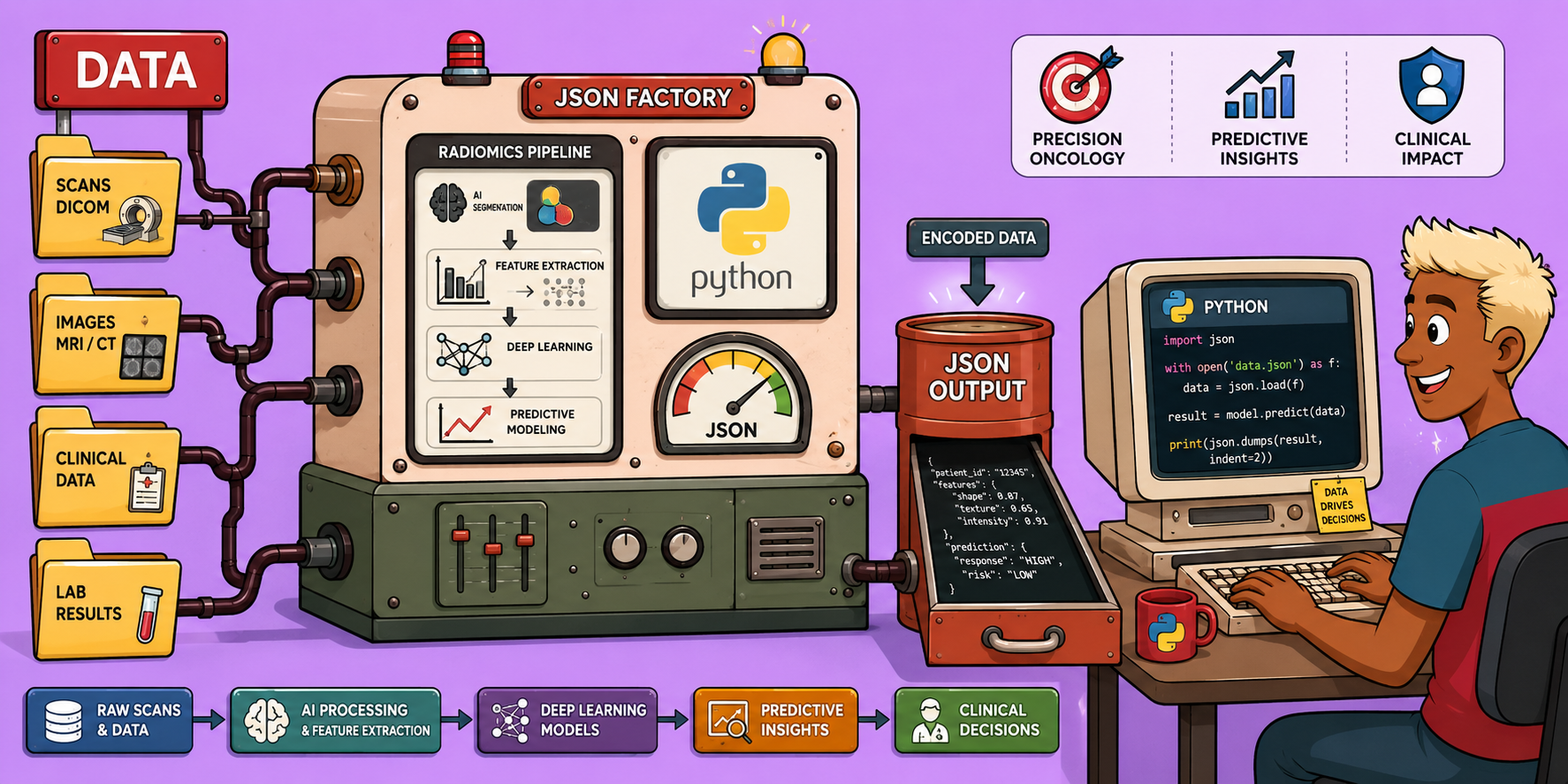
To harvest this immense amount of voxel data, every medical scan undergoes a rigorous six-phase blueprint within the radiomics engine:
- Acquisition: Obtaining the initial image data.
- Segmentation: Isolating the target of interest, such as a tumor.
- Preprocessing: Cleaning and normalizing the image data.
- Feature Extraction: Translating biological information into mathematical variables.
- Feature Selection: Refining the extracted features to identify the most predictive ones.
- Modeling and Validation: Building predictive models and confirming their effectiveness on unseen patient data.
Let’s delve into the crucial initial phases:
Phase 1: Segmentation – Finding the Needle in the Haystack
Accurate segmentation is paramount. If we aim to predict tumor behavior, we must perfectly isolate it. AI-driven auto-contouring is increasingly used to map tumor boundaries. Why? Because even a slight inclusion of healthy tissue within the boundary can corrupt the entire predictive model, rendering it useless.
Phase 2: Preprocessing – Cleaning the Dirty Lens
Before any mathematical extraction can occur, the data must be meticulously cleaned. This involves several steps:
- Normalization: Adjusting intensity values so scans from different hospitals or machines are mathematically comparable.
- Resampling: Ensuring all voxels are of the exact same physical size.
- Denoising: Applying filters to remove machine scatter and noise.
- Discretization: Grouping data to simplify subsequent analysis. This preparatory work is absolutely mandatory.
Phase 3: Feature Extraction – Translating Biology into Math
This is where the magic happens. From the cleaned and isolated tumor, algorithms extract hundreds of variables. These include:
- Shape metrics: Such as sphericity and compactness.
- First-order statistics: Like skewness and kurtosis, describing the distribution of intensity values.
- Complex spatial textures: Capturing patterns and variations within the tumor.
Together, these form a “feature barcode”—a highly unique mathematical fingerprint of the tumor. This mathematical texture is profoundly significant. For instance, a visually smooth tumor that is mathematically uniform might behave differently from a visually speckled, complex tumor. This disorganized mathematical texture often correlates with aggressive tumor mutations, treatment resistance, and poorer patient survival, effectively revealing biological severity through numbers.
Refining the Biological Signal: Overcoming Data Overload
Extracting thousands of features from a single tumor can lead to “data overload,” a phenomenon known as the “curse of dimensionality.” When you have thousands of features but only a small number of patients (e.g., 50 patients), the model can “overfit,” memorizing random noise rather than learning true biological patterns. This leads to data leakage and poor reproducibility.
To counteract this, data scientists act as “sculptors”:
- Principal Component Analysis (PCA): Condenses overlapping features into core components.
- LASSO Regression: Aggressively prunes irrelevant features, reducing their coefficients to zero.
This rigorous pruning leaves us with only the most robust and predictive variables, uncovering the true biological signal.
The Deep Learning Shift: A New Era of Analysis
We are currently witnessing a major paradigm shift in medical imaging: the transition from hand-crafted radiomics to deep learning.
- Hand-crafted Radiomics: Humans explicitly define geometric formulas (e.g., how to calculate entropy). These methods offer high interpretability and work well with smaller datasets but require significant manual effort for segmentation.
- Deep Learning: Neural networks process raw pixels through convolutional layers to automatically learn complex, invisible latent features that human mathematicians might never conceive. Deep learning offers raw, autonomous power with minimal human effort but requires massive datasets and operates as an opaque “black box.”
The future lies in hybrid models that combine the explainability of hand-crafted mathematical features with the immense power of neural networks, leveraging the best of both worlds.
Clinical Impact and Limitations: Bridging Theory and Reality
The impact of this technology is already tangible:
- Rapid AI auto-contouring: Significantly reducing planning time for physicians.
- Prediction of tumor control probabilities: Before treatment even begins.
- Generation of synthetic CTs: From MRI data, eliminating radiation exposure.
- Adaptive radiotherapy: Dynamically adjusting radiation beam pathways in real time as tumors shrink during treatment.
However, significant bottlenecks remain:
- Data drought: Medical data is often siloed due to privacy regulations.
- Standardization crisis: Scans from different manufacturers (e.g., Siemens vs. GE) can have mathematical differences.
- Black box dilemma: Doctors and regulatory bodies demand explainable AI. For life-and-death decisions (e.g., radiation dosage), understanding “why” an algorithm made a certain recommendation is critical.
Overcoming these challenges is the defining task for the next decade.
The Future of Oncology: Towards Personalized and Autonomous Care
The research frontier is expanding rapidly:
- Delta-radiomics: Tracking real-time biological treatment responses.
- Multi-omics: Fusing imaging features with DNA profiles for a holistic view.
- Federated learning: Training AI models across global hospital networks without sensitive patient data ever leaving its home institution, addressing the data drought.
All these advancements are converging towards fully automated, highly personalized care paradigms. We envision AI physicist assistants handling rigorous quality assurance, and massive foundation models adapting to medical physics tasks with minimal prior training. Ultimately, this trajectory points to real-time adaptive treatment, where autonomous radiation systems adjust instantly, guided by each patient’s unique, mathematically extracted digital twin.
Feature extraction is no longer just about image analysis; it is becoming the foundational language of personalized, predictive, and autonomous medicine. The provocative question for us all is: are we ready to fully trust the invisible mathematical language of our own biology as machines become increasingly adept at reading the invisible textures of disease?
🔍 Discover Kaptan Data Solutions — your partner for medical-physics data science & QA!
We're a French startup dedicated to building innovative web applications for medical physics, and quality assurance (QA).
Our mission: provide hospitals, cancer centers and dosimetry labs with powerful, intuitive and compliant tools that streamline beam-data acquisition, analysis and reporting.
🌐 Explore all our medical-physics services and tech updates
💻 Test our ready-to-use QA dashboards online
Our medical physics SaaS is coming soon — designed for radiation therapy, imaging, and nuclear medicine centers.
Our expertise covers:
🔬 Patient-specific dosimetry and image QA (EPID, portal dosimetry)
📈 Statistical Process Control (SPC) & anomaly detection for beam data
🤖 Automated QA workflows with n8n + AI agents (predictive maintenance)
📑 DICOM-RT / HL7 compliant reporting and audit trails
Leveraging advanced Python analytics and n8n orchestration, we help physicists automate routine QA, detect drifts early and generate regulatory-ready PDFs in one click.
Ready to boost treatment quality and uptime? Let’s discuss your linac challenges and design a tailor-made solution!
Get in touch to discuss your specific requirements and discover how our tailor-made solutions can help you unlock the value of your data, make informed decisions, and boost operational performance!
Comments